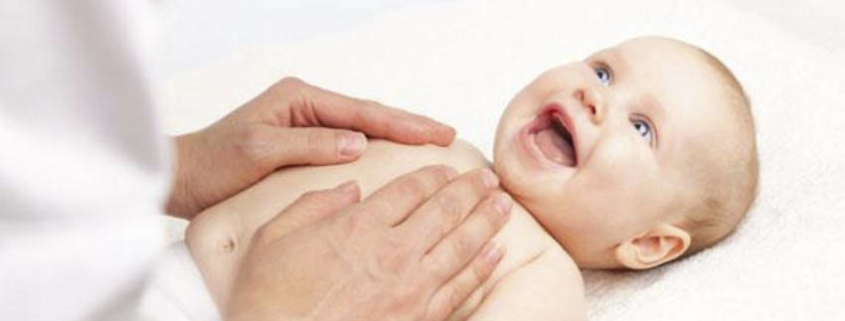
Osteopathy Treats Colic and Wind
Osteopathy Treats Colic and Wind – Mechanical stresses during delivery and pregnancy can aggravate or cause colic. Other factors that need to be considered are the mother’s diet, lactose intolerance, stress during pregnancy and a family history of colic.
The nerve to the stomach (vagus nerve) can be irritated as it exits at the base of the skull by birth compression. This retained compression on the nerve can reduce the efficiency of digestion and therefore impair its function.
Stress through the trunk of the baby as it passes through the birth canal, or from a poor first breath, or shock from the birth will increase torsion and/or tension within the diaphragm. Impairment of the diaphragm will compromise the ability of the stomach to hold on to and digest its contents.
Tension through the umbilical cord during delivery due to it being entwined around the baby’s neck can be spread to the diaphragm and gastrointestinal fascia. Thus the position and function of these areas can be disturbed.
Stress in the pregnant mother is also considered to increase the susceptibility of colic in the infant by making their nervous system more reactive.
A paediatric osteopath will be able to work on the restrictions within the baby’s body and effectively reduce the signs and symptoms of infantile colic.
The typical presentation of an infant with colic is abdominal distension, frequent gas emissions, apparent abdominal pain, irritability and excessive crying
Children with colic are often described as crying without identifiable cause and who are hard-to-soothe, although being otherwise healthy infants, well fed and showing no signs of failure to thrive.
Pathophysiology
Symptoms of colic reflect an increased excitability of the intestinal vagus nerve. (Cranial Nerve X). Altered (parasympathetic) function of the vagus nerve causes irregular stimulation of the intestinal muscles, producing spasms and constriction of the lumen in the intestine.
Above the constriction, the passage of the contents of the bowel is slowed. As a result of the digestive process gas develops which is not properly carried off. This results in a distension of the gut and colicky pains. If distension of the gut is prolonged, the musculature of the intestine becomes thinner than normal and its peristaltic power decreases, which is
The production of colicky pains is primarily due to parasympathetic over stimulation causing constriction; secondarily, due to dilation which irritates the sympathetic nervous system, which transfers the impulse to the spinal sensory nerves, producing the intestinal viscerosensory reflex.
Evidence In Practice
A study by Clive Hayden in 2006 showed osteopathy to be successful in the treatment of infantile colic (Hayden and Mullenger, 2006).
This study was based on 28 infants over a period of four weeks. They were split into two groups; one group receiving osteopathic treatment, and the other forming the control group. The infants who received osteopathic treatment showed a 63% reduction in crying and 11% increase in sleep, compared to only 23% reduction in crying and 2% increase in sleep in the control group.
This study therefore strongly suggests that osteopathic treatment can benefit infants with colic, however a larger double blind study is warranted.
How Osteopathy Helps With Infantile Colic
In newborns the head and neck generally bear the brunt of the mechanical forces encountered during labour and delivery., It is however designed to withstand these stresses and therefore most babies go on to be productive and well-adjusted adults.
The passage through the birth canal is a process for which most humans are extremely well adapted. The flexibility of the cartilaginous neurocranium produces a folding of the vault bones, similar to the arrangement of petals of a rose bud.
After delivery, the mechanical forces and movements associated with breathing, crying and suckling expand the cranium and correct the sutural overlap.
In some cases the normal forces of life are unable to resolve these strains, and we have a baby who may present with irritability, problems suckling, abnormal posturing, delayed postural patterning, or a myriad of other vague clinical symptoms.
Establishing a clinical diagnosis and understanding its aetiology are probably the most difficult tasks faced by a clinician caring for such an infant. As a result, ones index of suspicion must remain high for a broad differential diagnosis.
The head and neck play an important role in the establishment of posture and balance mechanisms. Strains and mechanical dysfunction may present immediately after birth, or with the expression of new developmental milestones.
Along with a thorough history, the symmetry and execution of new movement patterns can provide the osteopath with clues about somatic dysfunction in the infant.
Basic reflexes linking head and neck control with eye movements, mastication, suckling, vestibular activity, and control of the torso and extremities exist at birth. These influence and in turn are influenced by mechanical function in the craniocervical junction and the neck.
Osteopathic treatment is mainly directed at somatic dysfunction of occipitocervical junction and upper cervical spine for their effect of the vagus and parasympathetic somatovisceral reflexes.
Somatic dysfunction of the thoracic spine, ribs and upper lumbar spine may be treated to affect sympathetic somatovisceral reflexes. In other words, the organs involved in digestion are supplied by nerves emerging from the thorax (T5-T12). If these segments are dysfunctional by any means, the infant may present with digestion disturbances. Dysfunction in these areas also impacts the lymphatic and venous drainage of the abdominal contents, of which the diaphragm in extremely important.
Osteopthic treatment to these areas could therefore alleviate symptoms caused by these mechanical disturbances.
Sacropelvic somatic dysfunction may be treated to affect pelvic parasympathetic somatovisceral reflexes.
Occipital Bone At Birth
At birth, the occipital bone is complex. It is made up of four parts: a supraoccipital portion or squamous part, two lateral parts and the basiocciput.
The squamal and lateral parts are joined by a cartilaginous matrix. The lateral parts (masses) are joined to the basiocciput by cartilage as well. This is particularly relevant as cartilage is deformable.
Cartilage Is Deformable
You may test this hypothesis by pushing on your nose or ear.
The jugular foramen is located within the cartilage adjacent to the lateral masses.
Changes or deformations occurring within the basiocciput/lateral mass complex not only affect the shape of the occipital bone, but also affect the shape of adjacent structures and adjacent foramina.
Cranial Base Superior
From a clinical standpoint, that is very important, nerves and vascular structures do not pass through foramina in isolation. These structures are accompanied by venous plexuses, fascia and fat. The tendon and fascias of the pharyngeal and cervical musculature are attached to the external surface of the occipital bone.
Changes in the relationship of adjacent osseous structures will alter the relationship of the soft tissues structures attached to them.
This may create compression or stretch on the nerves and vessels passing in close proximity. Furthermore, alterations in tissue relationships may impede venous and lymphatic return from the area, leading to tissue congestion and compromising neurotrophic function.
Osteopathic Treatment
Osteopathic treatment involves the application of gentle manual techniques to the head as well as any other areas of the infant body that demonstrates palpable increased ligamentous/muscular tone, or decreased/abnormal articular mobility.
Very light tactile pressure is applied to the affected area until a palpable release of the relevant physical tensions and areas of dysfunction (including parts of the cranium) is achieved.
Example
Osteopathic treatment may alleviate the physical and biomenchanical influences of birth.
It is feasible that by reducing the distortions in the musculoskeletal framework, improving joint mobility, and reducing apparent muscular hypertonia in the infant, manipulation may reduce the somatic afferent load into the central nervous system.
Additionally, although colic may represent a ‘wastebasket’ of multiple aetiologies presenting with a similar picture, a plausible explanation for infantile colic in some children is that it is variant of cephalgia (headache).The common gastrointestinal manifestations such as nausea and vomiting that often accompany cephalgia may account for the GI symptoms we see in infants.
Disturbance of the Vagus nerve is a common accompaniment to cephalgia. The osteopathic literature includes much discussion of strain patterns common to colic. (Carreiro, 2003)
These include dysfunction in the cranial base, craniocervical junction, and upper and mid thoracic areas. These areas are also commonly involved in cephalgia.
In light of this we must consider that in some populations of infants the primary aetiology of the colic may be the somatic dysfunction in the head and neck, as it is in cervicogenic headaches.
In some children colic may also be a disease of the immature nervous system. An inability to screen out afferent input may lead to sensory overload, agitation and irritability.
The fact most children ‘grow out’ of colic at approximately the same time as they achieve developmental milestones in the brain and gut supports this argument.
The presence of somatic dysfunction in these infants may be a contributing factor, but not necessarily the primary cause of the child’s symptoms.
Lastly, colic may be a clinical manifestation of immature gut function, whereby the gut is responding to new antigenic materials. Regardless of the aetiology, there appears to be a commonly accepted involvement of the Vagal system. As such, anything that could be deemed as irritating to these systems should be addressed in an attempt to give the baby and the family some peace.
References
Carreiro, J.E. (2003) An Osteopathic Approach to Children, Elsevier Science Limited, China.
Hayden, C. and Mullenger, B. (2006) A preliminary assessment of the impact of Cranial Osteopathy for the relief of Infantile Colic. Complementary Therapies in Clinical Practice, Vol. 12, pp. 83-90
View a list of common complains that Osteopathy can assist with
Discovery the benefits of Osteopathy
- What is Osteopathy?
- Adult health issues
- Babies and Children
- During and after pregnancy
- Common Complaints
- Testimonials
- Sports Injuries
- Genral Osteopathy FAQs
- The Science & Reasearch


